
Lipoblastoma in an Infant
Case Report
Additional Files
Published
How to Cite
Issue
Section
License
Copyright (c) 2020 Juan Ramírez Pico, Edwin Edwin Ross Rodríguez, Mario Leone Pignataro, Andrés González Cabrera, Diana Alvarado Soto

This work is licensed under a Creative Commons Attribution-NonCommercial-ShareAlike 4.0 International License.
Keywords:
LIPOBLASTOMA, CHROMOSOME ABERRATIONS, INFANT, CASE REPORTAbstract
Introduction: Lipoblastoma is an infrequent benign neoplasm originating in adipose tissue, presenting almost exclusively in pediatric patients before the age of three years, predominantly in males. It is located mainly on the limbs and trunk, as a painless, progressively growing tumor. The treatment of choice is surgical and has a favorable prognosis. Recurrences occur in cases in which the resection could not be complete.
Clinical case: 7-month-old female infant. From the age of three months, he presented a progressive increase in the volume of the left lower limb. Physical examination revealed a large mass measuring 12 x 7 centimeters that involves the posterior aspect of the left thigh: a painless and well-defined mass. No compromise of mobility, no limb edema.
Diagnostic workshop: Magnetic resonance imaging reported a hyper-intense solid tumor of the left thigh in T1 and T2, hypo-intense in STIR with fine septa inside it, extending from the abductor magnus muscle of the biceps femoris and measuring 11.2 x 7.9 x 8.4 cm in its longitudinal, anteroposterior and transverse axes respectively, displacing and compressing the semitendinosus, semimembranosus and gracilis muscles. The patient underwent a complete and wide excision of the mass using a posterolateral approach.
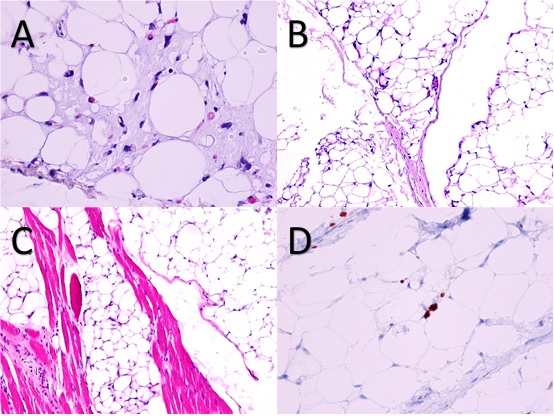
Outcome: Pathology reported a tumor made up of adipose tissue with myxoid tissue, without nuclear atypia; partial infiltration of the adjacent skeletal muscle is evidenced, without skin involvement, without necrosis and negative surgical margins for neoplasia. Immunohistochemistry with a cell proliferation marker KI-67, a positive result of 1%, and a negative MDM2 study (inhibitor of transcriptional activation of p53); findings consistent with lipoblastoma. The cytogenetic study was not performed.
Evolution: The patient was discharged on the fourth postoperative day without complications. With a complete recovery, in the fifth month of follow-up a new magnetic resonance study was performed in which no images that suggest residual tumor or tumor recurrence are visualized
Conclusion: Lipoblastoma should be taken into account as a differential diagnosis in children with soft tissue tumors, its treatment is eminently surgical with a good prognosis if the removal is complete.